

What Are The Differences Between The Livers Animals Comparative
Liver Topography (Pig) - Copyright Nottingham 2008
Introduction
The liver (hepar) is an extremely important organ in the body of mammals and vertebrates equally it provides functions essential for life. Information technology is the largest internal organ and has numerous functions including product of bile and protein, fatty and carbohydrate metabolism. During foetal evolution, the liver has an important haemopoetic function, producing scarlet and white claret cells from tissue between the hepatic cells and vessel walls.
The size of the liver varies due to its office in metabolism. In carnivores the liver weighs nigh 3-five% of body weight, in omnivores ii-three% and in herbivores 1.5%. the liver is much heavier in young animals than older animals as information technology atrophies with age.
The liver is derived from an outpocketing of endoderm epithelium on the ventral duodenum from the caudal part of the foregut. The connection to the gut narrows to become the bile duct. The parenchymal tissue of the liver is formed from proliferating epithelial cords or strands which integrate with the claret sinuses of the umbilical and vitelline veins. The mesoderm of the septum transversum forms the venous sinosoids and connective tissue of the liver.
Construction
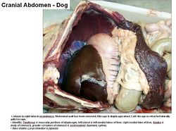
Topography of the Liver (Canis familiaris)- Copyright RVC 2008
The liver is located in the cranial part of the abdomen. Information technology is immediately caudal to the diaphragm and cranial to the stomach and intestines. Mostly the bulk of the liver is on the correct of the midline. Information technology is divided into lobes by fissures. Cranially the liver is convex, called the diaphragmatic surface. Caudally the liver is concave, chosen the visceral surface. The caudate lobe has a renal impression from the right kidney. The gastric impression occupies the whole of the left half of the visceral face. The duodenal impression at the junction of the right and quadrate lobes continues onto the right lateral and caudate lobes. Passages or notches on the median airplane allow the caudal vena cava and oesophagus to laissez passer by. The gall float is located betwixt the correct medial and quadrate lobes. Reticular fibres (collagen type III, proteoglycans and glycoproteins) back up the hepatocytes and walls of the sinusoids. Interlobular spaces support bile ducts and blood vessels. The lesser omentum (often fat filled) is on the visceral surface between the left lateral lobe, heptic porta and lesser curvature of the breadbasket. In that location is a oesophageal notch where the oesophagus passes over the liver.
Divisions of the Liver
The liver can be divided into lobes, lobules, hepatocytes and sinusoids.
Lobes of the Liver
The lobes of the liver include the left lateral, left medial, right lateral, right medial, quadrate, caudate and papillary.
Ligaments
The coronary ligament attaches the liver (from the diaphragmatic surface) to the diaphragm. It is an irregular fold of peritoneum. Information technology surrounds the triangular base of the diaphragmatic surface. It is continuous with outer most layer of the caudal vena cava. The falciform ligament is ventral to the coronary ligament. It is a fat filled embryological remnant of the fetal blood vessels from the placenta. It causes problems for surgical entry into the belly. It is located cranial to the umbilicus and is a vestige of the umbilical vein. The triangular ligament is on the right and left sides of the coronary ligament.
Office
Production of bile run across Bile Germination .
Nearly all the blood circulated effectually the belly flows dorsum through the portal vein to the liver where it comes in contact with the liver cells, ensuring the products of digestion are presented to the hepatic cells earlier inbound the general circulation. Other functions include carbohydrate metabolism, glycogenesis, glyconeolysis, gluconeogenesis and the breakup of insulin and other hormones. Protein metabolism produces soluble mediators of the clotting cascade, Albumin and hormone transporting globulins. The liver is also involved in lipid metabolism, lipogenesis and the synthesis of cholesterol.
The liver has a function in hormonal command of the following; Insulin and glucagon, Glucocortocoids, Catecholamines and the synthesis of other important hormones (come across Endocrine Arrangement). It also has a role in immunoregulation via kupfer cells and the complement synthesis and metabolism.
The liver is important in storage of water soluble vitamins, fat soluble vitamins, fe, triglyceride and glycogen.
The liver breaks down haemoglobin and toxic substances through drug metabolism. It converts ammonia to urea and allows the direction of endogenous waste, e.g haem (Hb, cytochromes, Mb) and ammonia (amino acids).
Vasculature
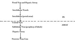
Blood Flow in the Liver - Copyright nabrown RVC 2008
The liver has a dual blood supply. 70-fourscore% via the hepatic portal vein (nutrient rich) and 20-30% via the hepatic artery (oxygen rich). It has a big blood supply (most a i/three of cardiac output passes through the liver).
The hepatic artery is a co-operative of the caeliac artery. The portal vein is formed past tributaries draining the spleen, pancreas and digestive tract. Intrahepatic arteries combine with portal vein branches to supply the connective tissue and hepatic sinusoids of the liver. Claret flows from the portal areas into the fundamental vein. The fundamental vein is lined by uncomplicated squamous epithelium. The bile duct, claret vessels (including the important hepatic vein) and nerves enter and go out the liver at the hepatic porta. Blood from the central vein opens into the caudal vena cava. Liver circulation is controlled by interarterial, intervenous, arteriovenous, and by sphincter mechanisms, allowing careful regulation.
Innervation
The liver is innervated by sympathetic nerves from the periarterial plexuses and parasympathetic nerves from the vagal body.
Lymphatics
Efferent vessels pass to hepatic nodes around the hepatic porta. The lymph drains into the visceral cysterna chyli. Some lymph travels to the accessory hepatic and caudal mediastinal lymph nodes on the caudal vena cava.
Hepatic Duct Systems
There are canaliculi within lobules. Canaliculi open up into larger ductules then into a few large hapatic ducts. Before and before long subsequently leaving the hepatic porta, the ducts combine into a single torso which runs to the duodenum. The cystic duct runs from common trunk to the gall float transporting bile from the liver to the gall float. Distal to the cystic duct is the bile duct (ductus choledochus) which transports bile from the gall bladder into the duodenum. There are no valves, and then bile may flow in either direction.
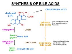
Germination of Bile Acids - Copyright RVC 2008
Bile Acids
Bile acids are equanimous of cholesterol, bile acids and steroids. The chief bile acid is cholic acid (C24). Conjugated to taurine or glycine in the liver to reduce pKa so they exist in an ionised form as bile salts. Bile salts cohabit with cholesterol and phospholipids and are then secreted into the bile. They emulsify fats which helps absorb fat soluble vitamins. In aqueous solution, they class micelles which are amphiphilic and can transport free fatty acids beyond the castor border.
Species Differences
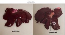
Liver Topography (Dog) - Copyright Nottingham 2008
Canine & Feline
Both the left and correct lobes are subdivided. Complete obstacle of the hepatic artery is fatal. The liver is about entirely intra-thoracic. An enlarged caudate process contacts the right kidney.
Equine
The liver is contained entirely within the rib cage, to the right of the midline. It is less lobated. There is no gall bladder and the left lobe is subdivided. At that place is no papillary lobe. In the foal, the liver is larger and more symmetrical. The bile duct opens into the duodenum at the aforementioned papillae as the major pancreatic duct. Bile is constantly secreted.

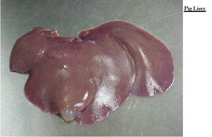
Liver Topography (Squealer) - Copyright Nottingham 2008
Porcine
The liver has deep interlobular fissures and a large amount of interlobular connective tissue. It has a mottled advent. A Deep interlobular fissure divides the liver into 4 lobes- the left, right, medial and lateral. There is a modest caudate lobe (which does not contact the kidney and so no renal impression). It is mostly on the right of the midline and has no papillary lobe.
Ruminants
The liver is entirely displaced to right of the midline. It has fused lobes.
Small Ruminants Sheep accept a deeper umbilical fissure than cows. Sheep also accept a smaller caudate lobe than cows and accept two papillary processes.
Avian
Encounter avian liver
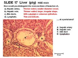
Histology
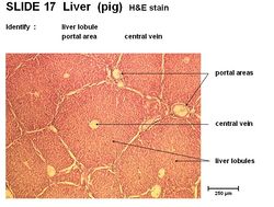
The larger liver cells are called lobules. Each lobule contains an opening for the primal vein and contains portal areas. The lobules are composed of liver cords chosen hepatocytes. Sinusoids are present between hepatocytes containing red blood cells. There is a connective tissue sheathing around each liver lobule. A thin mesothelium covers the connective tissue layer.
The portal expanse present in the lobules contains the hepatic artery, which has thick walls and a small-scale diameter and the hepatic vein, which has thin walls and a large and irregular shape. It also contains bile ducts, with cuboidal or columnar epithelium and lymphatics that are small and frail.
Hepatocytes are the smaller liver cells in the lobules. They incorporate glycogen granules and take a spherical nucleus. They form cords called branching plates (lamellae). The upper and lower margins are tight junctions. They have 3 performance surfaces. Kupfer macrophages are nowadays nearly the lining of the sinusoids. The hepatocytes stain pink as they are eosinophilic.
Acinus
- the smallest functional unit of the liver - this unit of measurement stresses the dependence of the liver on its afferent blood vessels and efferent bile ducts
- a roughly diamond shaped parenchymal mass surrounding and supplied by the portal expanse
- consists of three zones:
- Zone 1 - periportal (centroacinar), around the portal areas
- Zone two - midzonal
- Zone three - centrilobular (periacinar), adjoining the hepatic venules
NB: blood enters from Zone 1 to Zone 3 and thus becomes less and less oxygenated
Links
Click here for data on pathology of the Liver
Click hither for information on Hepatic Stellate Cells
Click here for information on Bile Formation
Click here for data on portosystemic shunting
| Liver - Anatomy & Physiology Learning Resources | |
|---|---|
| To reach the Vetstream content, please select | Canis, Felis, Lapis or Equis |
| Test your knowledge using elevate and driblet boxes | Compare liver structure in different species |
| Examination your knowledge using flashcard type questions | Liver - Anatomy & Physiology - Flashcards |
| Choice of relevant videos | Bovine liver potcast Bovine liver potcast 2 Ovine liver with hepatitis cysticercosa potcast Equine liver with hydatid cysts potcast Equine liver potcast Liver lobes of the Horse Liver lobes of the Cow |
| Pick of relevant PowerPoint tutorials | Histology of the liver - see part 2 |


Source: https://en.wikivet.net/Liver_-_Anatomy_%26_Physiology
Posted by: perrywhearommens.blogspot.com
0 Response to "What Are The Differences Between The Livers Animals Comparative"
Post a Comment